The ALARA+ Coalition formed after the Summit on Radiation and Orthopedic Risks in Fluoroscopic Laboratories at SCAI Scientific Sessions 2025. The member organizations have come together to fulfill a common vision; to end common injuries to healthcare professionals by increasing the adoption of modern protective strategies and technologies.
Best practices for reducing harmful effects
Managing radiation exposure
- Equipment Optimization. Using the lowest appropriate frame rate and dose settings.
- Real-Time Awareness. Monitoring doses during procedures and adjusting techniques.
- Time. Minimize radiation exposure duration, using lower frame rates when appropriate rather than default higher rates.
- Distance. Maximize distance from the radiation. Use extension devices and remote controls when possible and step away from the table when not operating.
Fundamentals
Radiation protection has two components—passive (protective equipment in the lab) and active (proper user of equipment, staff training, dose monitoring, and radiation-reduction techniques) processes6.
Active/Passive Radiation Techniques & Devices7
Active Processes
- Education and training of the staff
- Routine radiation dose monitoring
- Personal dose meters
- Real-time dose monitoring
- Procedural techniques in reducing radiation exposure
- Limiting fluoroscopy time
- Minimize use of high contrast modes
- Use of lower frame rates whenever possible
- Avoiding use of steep angulations
- Utilize available radiation-reducing technology
- Virtual collimation
- Last image hold
- Storage of fluoroscopy
- Low pulse-rate fluoroscopy options
- Low dose-per-frame
- Low frame rate options
- Spectral beam filtration
- Higher X-ray beam energy
- Use of image noise reduction technology
- Distance from radiation source
- Optimal table positioning-higher table setting if possible
- Staying at low scatter radiation areas for the staff
- Keeping non-target anatomy away from the X-ray beam
Passive Processes
- Architectural shielding
- Rolling leaded transparent shields
- Stationary leaded transparent shields
- Equipment-mounted
- Ceiling-suspended shields
- Table-suspended curtains and drapes
- Radioabsorbent patient drapes
- Radial arm boards
- Personal protective equipment
- Caps
- Eyewear
- Thyroid collar
- Aprons
- Lead acrylic face mask
Shielding includes architectural (built into fluoroscopy practice structure, including rolling and stationary leaded shields8, equipment-mounted, and personal protective devices9.

Radiation protection equipment in a modern cardiac catheterization laboratory. In this picture you will find ceiling-mounted lead glass shield, under-table lead curtain, radioabsorbent patient drape, real-time radiation dose monitor, Zero-gravity system, and a rolling lead shield.
Best practices
- Exercise. Regular exercises focused on core strengthening, postural training, and flexibility to counter the strain of wearing heavy protective equipment.
- Protective measures. Proper shielding protocols, optimized equipment positioning, reduced fluoro time, and appropriate use of protective barriers.
- Interpreting dosimetry reports. Terms to know:
- DDE = Deep dose equivalent (1cm depth-deeper tissues)
- SDE = Shallow dose equivalent (0.007cm deep - skin level)
- LDE = Lens dose equivalent (eye lens radiation dose)
- Exposure reduction rules. Evidence-based protocols to minimize radiation dose to both patients and staff.
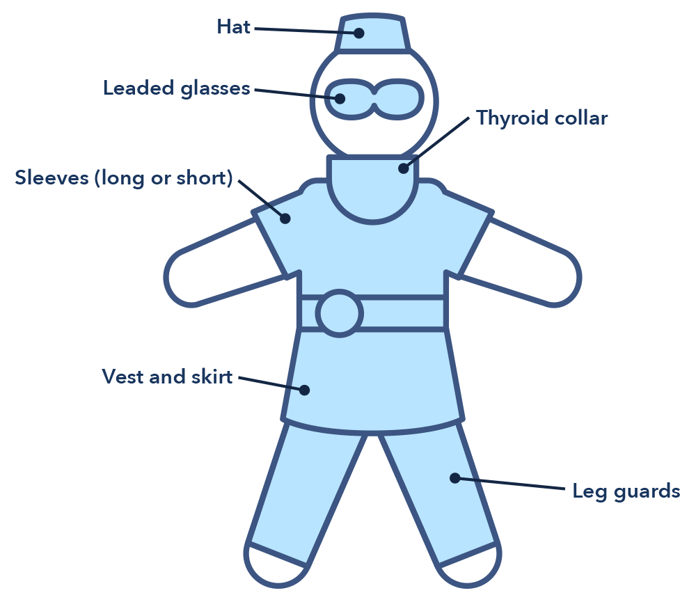
Figure of full coverage outfit:
- Apron Thickness Standards. 0.5 mm lead stops 95% of scatter radiation.
- Thyroid Protection. Thyroid shields should always be worn—it is a radiosensitive organ and cancer risk exists despite age of exposure.
- Eye Protection. Leaded glasses reduce eye radiation by 35 to 90%; lead acrylic face masks can reduce exposureby up to 97%10.